The spontaneous dynamics of the brain of the zebrafish is close to a critical point
The spontaneous dynamics of the brain of the zebrafish is close to a critical point
Such is the conclusion of a study published on 15 November in the journal Neuron by a multidisciplinary, international team of researchers, of which Adrián Ponce-Álvarez, a member of the research group in Computational Neuroscience, is the first author, with the participation of Gustavo Deco, ICREA of the DTIC and director of the Center for Brain and Cognition, in collaboration with researchers of the Institute of Biology of the École Normale de Paris (IBENS).

Just as condensed matter can be ordered (e.g., ice) or disordered (e.g., water vapour), the collective activity of neural networks can also experience phase transitions between order and disorder. These phase transitions are separated by critical points in which order and disorder coexist. The mixture of order and disorder means that the repertoire of patterns of neuronal activity is maximal and produces emerging dynamic patterns on all spatial and temporal scales. For this and other reasons, previous studies have suggested that criticality optimizes various brain functions.
The authors of a new study published on 15 November in the journal Neuron have observed the in vivo dynamics of the whole brain in zebrafish larvae with cellular resolution. Research carried out by and international, multidisciplinary team of researchers of which Adrián Ponce-Álvarez, a member of the research group in Computational Neuroscience, is the first author, with the participation of Gustavo Deco, ICREA of the DTIC and director of the Center for Brain and Cognition, in collaboration with researchers of the Institute of Biology of the École Normale de Paris (IBENS).
This study is the first to investigate criticality in an intact nervous system, in vivo, in the three dimensions of space and with cellular resolution
This study is the first to investigate criticality in an intact nervous system, in vivo, in the three dimensions of space and with cellular resolution. In addition, the authors warn that it has not been studied in depth how the environment affects this critical state, nor which are the mechanisms that cause functional connectivity to lead to this critical point.
Neuronal activity recorded using the light-sheet fluorescence microscopy technique has been analysed by theoretical neuroscientists at Pompeu Fabra University, using tools and concepts of Statistical Physics
To observe the dynamics in vivo of the whole brain, the researchers at the Institute of Biology of the École Normale de Paris (IBENS) have used one of the most promising new microscopic techniques currently in existence in the fields of cell biology, developmental and systems biology: light-sheet fluorescence microscopy. This technique enables observing the cellular activity of hundreds of thousands of neurons in transgenic fish larvae that express a fluorescent molecule indicative of calcium fluctuations (indicators that are in turn of cell activity). Neuronal activity thus recorded has been analysed by theoretical neuroscientists at Pompeu Fabra University, using tools and concepts of Statistical Physics.
“Neuronal avalanches show scale invariance, i.e., the statistics of the avalanches is similar at different scales of magnitude, time and frequency”
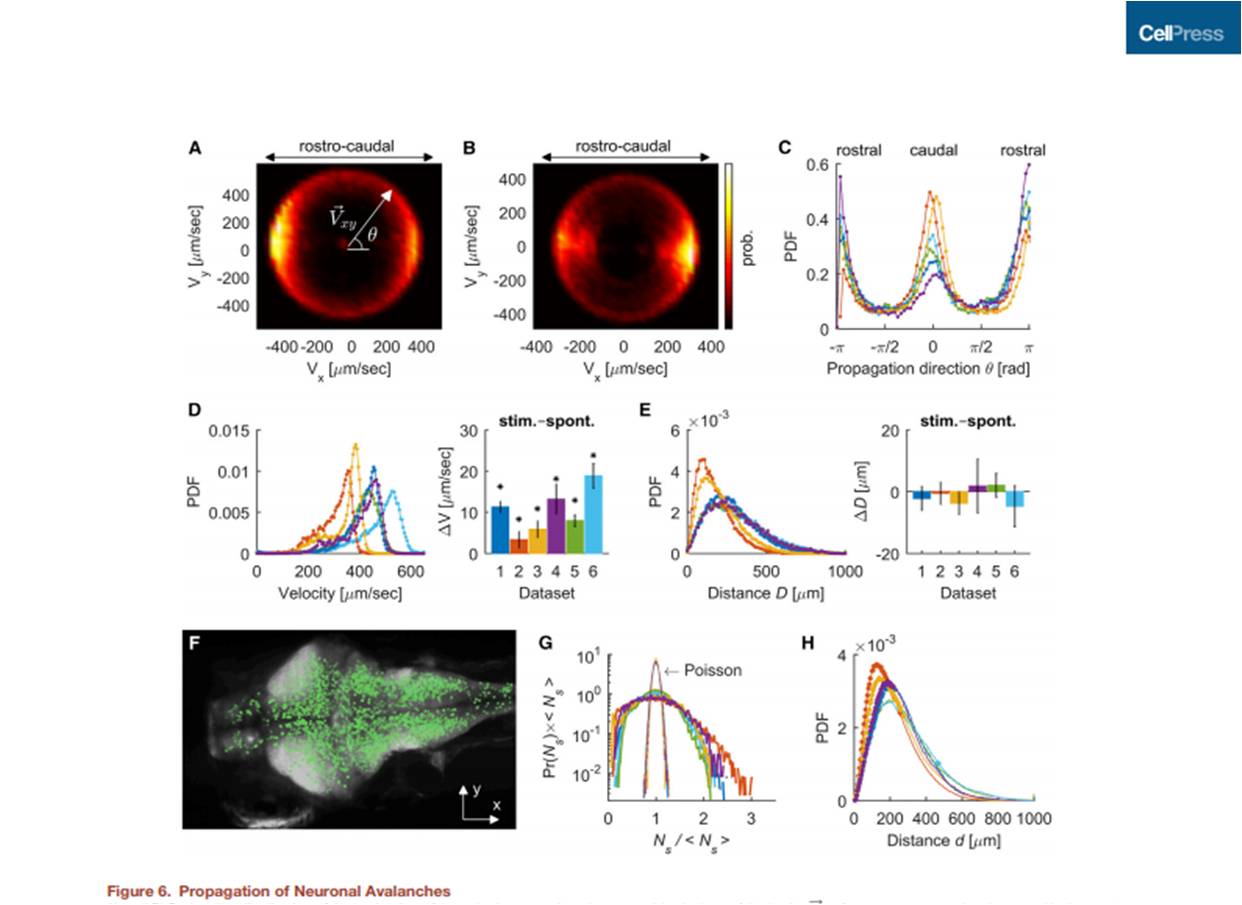
With this new technique “we can observe that the spontaneous activity of the brain is spread in three-dimensional space generating cascading collective events, or neuronal avalanches, that show scale invariance, i.e., the statistics of the avalanches is similar at different scales of magnitude, time and frequency”, says Adrián Ponce-Álvarez, first author of the article.
“In addition, we have found that by pharmacologically disrupting the coupling between neurons, the nervous system moves away from criticality and its ability to transmit information decreases”
The authors observed neuronal avalanches that propagated in the brain during periods of spontaneous activity induced visually. They analysed the spatiotemporal statistics of the neuronal avalanches and compared these statistics in various situations: during spontaneous activity, during the presentation of visual stimuli, during self-generated motor behaviours and during slight pharmacological disturbances. “Our results suggest that the spontaneous dynamics of the entire brain fluctuates near the critical point of a phase transition, but, when the system interacts with its environment (through sensory stimuli or self-generated movements), the dynamics can move away from the critical point. In addition, we have found that by pharmacologically disrupting the coupling between neurons, the nervous system moves away from criticality and its ability to transmit information decreases”, explains Ponce-Álvarez.
In conclusion, the authors have shown on the basis of an in vivo model and at cellular level that the central nervous system remains spontaneously in a critical state with scale invariance. However, in its interaction with the environment (sensory stimuli and self-generated inputs), the brain moves towards a more ordered regime, with less diversity of activity patterns, perhaps to restrict the possible sensory and motor representations and thus select the most appropriate.
Reference article:
Adrián Ponce-Álvarez, Adrien Joaury, Martin Privado, Gustavo Deco, German Sumba (2018), “Whole-Brain Neuronal Activity Displays Crackling Noise Dynamics”, Neuron, 15 November, DOI: 10.1016/j.neuron.2018.10.045.
