A model of the left ventricle might help better understand some cardiomyopathies
Made by members of the PhySense research group coordinated by Bart Bijnens, ICREA research professor with the Department of Information and Communication Technologies, published in the International Journal for Numerical Methods in Biomedical Engineering.
In the development of many species, most of the heart muscle is a spongy mesh of interwoven myocardial fibres. In mammals, as normal development progresses, the amount of these trabeculated structures decreases. This process is particularly evident in the ventricles, especially the left ventricle.
One of the hypotheses of trabecular development suggests that the formation of the trabecula is an initial step in the formation of the myocardium that allows accumulating a large amount of tissue temporarily perfused by the surrounding blood. More recent studies on the growth of myocardial cells suggest that once the trabeculations are formed, cell proliferation concentrates on the outside of the wall of the myocardium, forming a compact layer.
The interruption of this compaction process might explain the persistence of the trabeculae and the appearance of a double layer of the myocardial wall. A non-compacted left ventricle is a disorder of the myocardium characterized by increased trabeculation of the chamber of the left ventricle, that is, a two-layer myocardium with a thin, compact subepicardial layer and a thicker, non-compacted hypertrabeculated layer. NCLV is a condition that may present itself with varying degrees of ventricular dilation and dysfunction.
The relationship between trabeculation and heart function
Still little is known about the clinics of the interior structure of the heart chambers. The growing awareness of non-compacted cardiomyopathy has aroused the need to find out how the ventricular wall is organized in order to understand the role of myocardial trabeculation in the healthy heart.
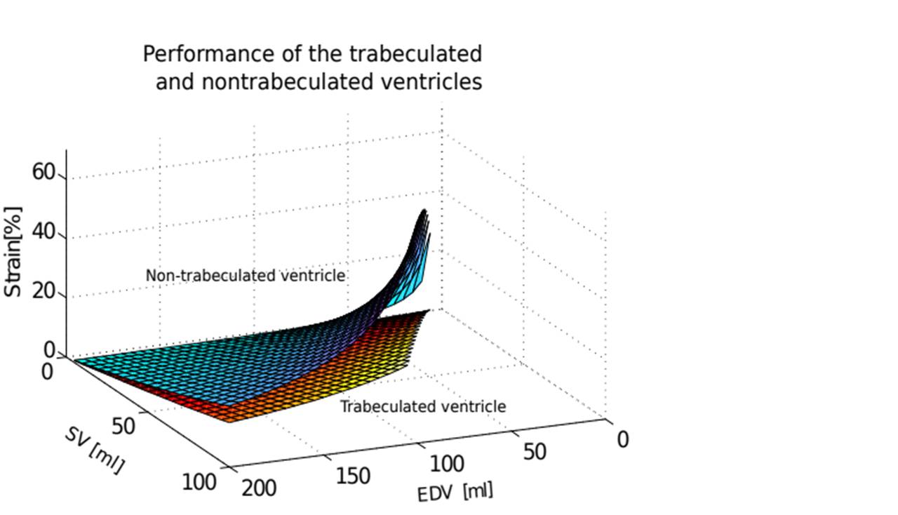
A study published in the advanced online edition of the International Journal for Numerical Methods in Biomedical Engineering has studied the relationship between trabeculations and heart function and has reached the main conclusion that they may serve to help the heart to increase its stroke volume without increasing the deformation of the organ.
This study has been carried out by Bruno Paun and Constantine Butakoff, members of the PhySense (Sensing in Physiology and Biomedicine) research group coordinated by Bart Bijnens, co-author of the article and ICREA research professor with the Department of Information and Communication Technologies (DTIC) at Pompeu Fabra University.
An ellipsoidal representation of the left ventricle
In the study, the authors present an ellipsoidal representation of a left ventricle, a theoretical analysis using non-invasive modelling techniques that allows a greater understanding of the function of the trabeculae in the generation of the cardiac output, for different sizes of the ventricle.
In this modelling, the trabeculations are represented through the fixed volume they occupy (without a predetermined form). Ventricular contraction is modelled by scaling the ellipsoid whereupon the measurements of longitudinal strain, end-diastolic, end-systolic and stroke volume are derived and compared. The authors’ main conclusion is that the trabeculations can be used to help the heart to increase its stroke volume without increasing its deformation and an optimal amount of trabeculations can be established from the point of view of deformation.
The trabecular pattern varies with age, gender; in athletes and during pregnancy
It is known that trabeculations are more frequent among young people and that they decrease with age, and that they present differences according to gender. For example, young athletes show a higher prevalence of left ventricular trabeculation and this has been seen even more among athletes of African/Afro-Caribbean origin.
The authors demonstrate that a trabeculated ventricle can work with less deformation, in comparison to a non-trabeculated ventricle, to produce the same stroke volume, which might explain why athletes and pregnant women develop reversible signs of non-compaction of the left ventricle, quite possibly because the trabeculations can increase cardiac output. Thus, this knowledge may help to evaluate patients with heart failure and with dilated cardiomyopathies that often show signs of non-compaction.
Reference article:
Bruno Paun, Bart Bijnens, Constantine Butakoff (2017),"Relationship between the left ventricular size and the amount of trabeculations", Journal for Numerical Methods in Biomedical Engineering, published online, 9th of november, doi: https://doi.org/10.1002/cnm.2939.