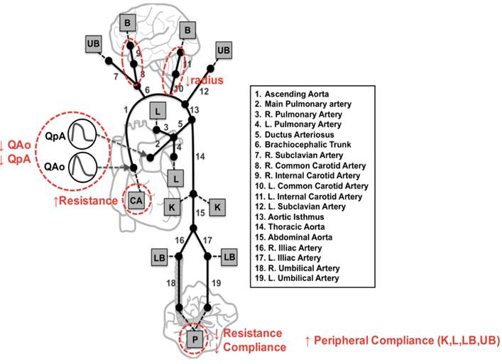
The blood flow of the foetuses of diabetic mothers is redistributed
These are the findings of a study that used computational modelling and the simulation of foetal circulation by the Physense research group coordinated by Bart Bijnens. The results were presented at the European Cardiology Society’s European Association of Cardiovascular Imaging, held from 7 to 10 December 2016 in Leipzig (Germany).
It is known that gestational diabetes mellitus (GDM) increases the risk of hypertension in the offspring. Often the foetuses of mothers with the disease (FGDM) have structural and functional myocardial abnormalities. However, it was not known whether cardiovascular circulation is altered in these foetuses as a result of GDM.

Hence, the main goal of a study coordinated by Bart Bijnens, ICREA research professor with the Department of Information and Communication Technologies (DTIC) and head of the Physense research group at UPF, and Eduard Gratacós, head of the Maternal and Foetal Medicine Service at Hospital Clínic de Barcelona, was to explore the cardiovascular circulatory patterns of the foetuses of mothers with diabetes mellitus. The results of the work were presented at the annual meeting of the European Cardiology Society’s European Association of Cardiovascular Imaging (EACVI), which was held from 7 to 10 December 2016 in Leipzig (Germany).
Babies born of mothers with diabetes tend to be larger, and their placenta is bigger, especially if the diabetes is not well controlled. There are data to suggest that some other organs such as the pancreas and kidneys could be affected. “We know that gestational diabetes mellitus affects the foetal organs”, said Aparna Kulkarni, paediatric cardiologist of the Bronx-Lebanon Hospital Center, New York, USA and first author of the study. Previous investigations have identified subclinical changes in the heart muscle of the foetuses of diabetic mothers.
In the paper presented at the EACVI, the researchers measured foetal blood flow using Doppler echocardiography. More specifically, they measured the blood flow of the brain, the left and right heart outflow tracts, the aorta, and the placenta. With the data obtained they built a computational model that simulates all the foetal circulation. This model was developed by Patricia García-Canadilla at the Physense research laboratory coordinated by Bart Bijnens, co-author of the study, at Pompeu Fabra University.
The research revealed that in comparison with the control group foetuses, in foetuses of diabetic mothers more blood flows to the placenta and less to the cerebral arteries, as well as a lower cardiac output. The researchers’ interpretation of this is that the foetuses of diabetic mothers receive a greater blood supply because they have changes in their blood vessels, although at the expense of irrigating the brain to a lesser extent, which is an interesting finding that could have greater implications.
This study was carried out with the participation of 14 foetuses of mothers with type 1 or 2 diabetes, and 16 foetuses of mothers without diabetes (control group). Nine of the mothers with diabetes used insulin, three took oral drugs, and two only dieted to control their glucose levels.
As the authors of the study state: “The placenta, once the baby is born, is no longer part of the circulation, but it is possible that the reduced blood flow to the brain that the foetus has received while in the uterus may affect the baby throughout its life. We do not know why this redistribution of blood flow takes place or the implications that this entails. Therefore, more research is needed to find out if it has a long-term impact on the health of the baby and if anything can be done to prevent it”.
Reference work:
A Kulkarni, P Garcia Canadilla, A Khan, K Beckerman, F Crispi, M Cruz-Lemini, B Valenzuela Alcaraz, O Gomez, E Gratacos, B Bijnens (2016), “Altered circulation in foetuses of diabetic mothers: a foetal computational model analysis”, Abstract P1267, Session no. 6: Cardiomyopathies, December 10, EuroEcho-Imaging 2016, 7-10 December, Leipzig (Germany). European Heart Journal Supplements ( 2016 ) 17 ( Supplement 2 ), ii269.
