Quantitative computational tools to interpret electro-anatomical maps in cardiology
A study coordinated by Oscar Camara, researcher with the Physense research group, published in the IEEE Journal of Translational Engineering in Health and Medicine that provides solutions for the analysis of cardiac electrical patterns.
The acquisition of a patient’s clinical data includes a set of techniques and procedures to obtain signals and images of the human body for clinical purposes, for diagnosing diseases, and scientific ones, for the study of normal anatomy and its function.
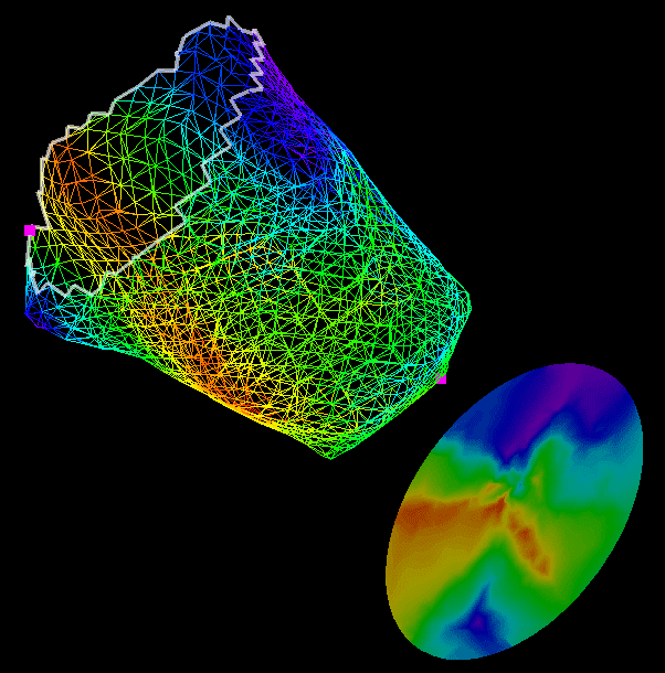
Electro-Anatomical Maps (EAM) are a type of medical data that fuse anatomical and electrical information and are commonly used in cardiology for the analysis of electrical activation in specific diseases. They provide detailed three-dimensional information of the voltage and activation time and act as a guide in interventions such as radiofrequency ablation in complex arrhythmias.
The main goal of a study conducted by researchers from the Physense (Sensing in Physiology and Biomedicine) research group, coordinated by Oscar Camara, a researcher with the Department of Information and Communication Technologies (DTIC) at UPF, is to find procedures to evaluate changes in the electrical activation of the heart in a patient at different moments in time, and differences between different individuals, helping to better understand the pathophysiological mechanisms that take place in heart disease.
The study, published on 6 January 2017 in the IEEE Journal of Translational Engineering in Health and Medicine, has involved other members of the Physense research group: David Soto Iglesias, as first author, Constantine Butakkof and Bart Bijnens (ICREA), together with researchers from the Cardiology Department at Hospital Clínic of Barcelona and the Asclepios research project of Inria Sophia-Antipolis in France.
Electro-anatomical maps provide an anatomic mesh of electrical activity
Electro-anatomic maps are constructed by processing data from intracardiac electrograms located in such a way as to provide an anatomic mesh made from a discrete set of points for which electrical and anatomical information is available. This information is used to carry out qualitative analyses of the changes that occur in cardiac electrical activity patterns in different conditions, such as in the case of a left bundle branch block, or in the case of a cardiac resynchronization device, or pacemaker, implantation.
However, electro-anatomical maps are difficult to compare, since they are extremely dependent on several factors such as patient anatomy; the number, locations and accuracy of the points used to acquire data, etc. Overcoming these constraints is a huge challenge and the work by Oscar Camara et al. provides solutions for the analysis of the electrical patterns of patients’ electro-anatomical maps.
A method that analyses the electrical activation patterns of the anatomic mesh
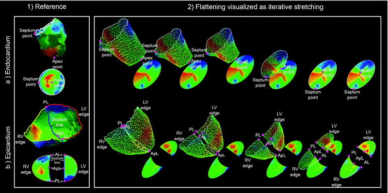
The first contribution of this study is the development of an automatic two-dimensional representation of the behaviour of the left and right ventricles of the heart obtained from three-dimensional electro-anatomical mapping.
The second contribution is the creation of a set of indices derived from the cardiac electrical activation: electrical wave speed and propagation, histograms to analyse local electrical differences in the ventricles, etc., which the researchers have applied in an experimental model to identify the physiological mechanisms underlying left bundle branch block and the analysis of treatment by means of cardiac resynchronization.
To illustrate the benefits of this procedure, the authors have created a set of controllable synthetic activation patterns on which to make the necessary representations and validate the indices and the proposed model. The authors have analysed and compared electrical activation patterns in a porcine model, specifically on four animals, two without any disease, and two with induced myocardial infarction with a cardiac resynchronization device implanted. The results confirm certain assumptions regarding the optimal location of the electrodes in devices for cardiac resynchronization.
Reference work:
David Soto Iglesias, Nicolas Duchateau, Constantine Butakoff, David Andreu, Juan Fernández-Armenta, Bart Bijnens, Antonio Berruezo, Marta Sitges, Oscar Camara (2017), “Quantitative Analysis of Electro-Anatomical Maps: Application to an Experimental Model of Left Bundle Branch Block/Cardiac Resynchronization Therapy”, IEEE Journal of Translational Engineering in Health and Medicine, doi.: 10.1109/JTEHM.2016.2634006.