Scientists show in 4D-imaging how neuronal and sensory cells form in the ear

Our ears, eyes and other sensory organs collect information about the world around us. Their formation during embryonic development is a complex process of which we still have much to learn, since the understanding of how the differentiation and specialization of cells in space and time are controlled is still a major challenge for biology. A study published in the journal eLife provides 4D images of the embryonic development of the inner ear in zebrafish, providing a new insight into the formation of this organ.
The inner ear is responsible for balance and hearing and its specialized cells, known as hair cells, detect sound and position. This information is passed on to other cells called sensory neurons, which relay the information to the brain. Both of these cell types originate from a pool of “progenitors” located in an embryonic structure called the otic vesicle, where the progenitor cells follow different sets of instructions to make hair cells or sensory neurons. The interpretation of this information results in the activation or repression of several genes important for the final destiny of these cells.
By using high resolution imaging techniques, the scientific team led by Cristina Pujades, researcher at the Department of Experimental and Health Sciences(DCEXS) at UPF, has reconstructed the dynamic event histories of individual hair cells and sensory neurons in the inner ear of zebrafish embryos within their native context. The combination of 4D microscopy with other imaging tools has allowed them to create a dynamic map of the progenitor cells of the otic vesicle.
4D microscopy includes the temporal magnitude, hence it is so useful for embryonic development studies: in addition to visualizing the samples in 3D format (with volume), the movements of the cells in time are recorded. “We have been able to perform spatiotemporal measurements, and thus to understand for the first time the behaviour of progenitor cells, that is, how their final destiny is related to their behaviour at a particular time and place”, says Sylvia Dyballa, the first author of the article.
The importance of spatiotemporal position in development
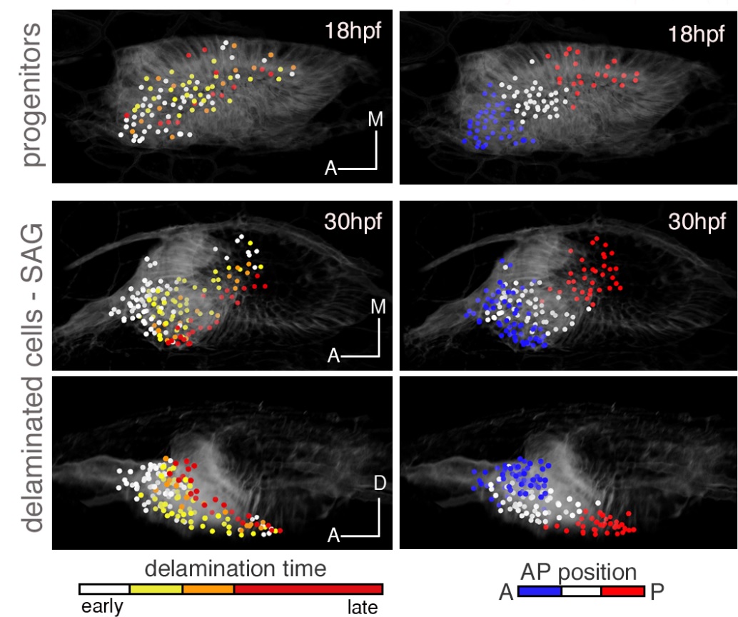
One of the processes that the team could observe in detail was the delamination of the neuronal progenitor cells. These cells are found in the otic vesicle; they depart from this structure by delamination and form the neural ganglion. The researchers observed that the organization and function of the sensory neurons in the ganglion depended on the behaviour of their progenitors during the delamination process.
“All these findings establish a link between the place and order of the progenitors’ delamination and their neural identity within the ganglion, and suggest the existence of fine spatial and temporal regulation in the embryonic development of the inner ear”, says Pujades., leader of the Research Group in Developmental Neurobiology.

Many changes in a short period of time
Delamination occurs massively in a very short period of time. Therefore, the group of neural progenitors undergoes dramatic changes in size and shape while keeping the homeostasis of the system. In addition, this process has to be coordinated with the formation of hair cells, which are in the first instance in contact with the outside. The researchers observed that these cells' progenitors have varying behaviours but share the fact that their birthplace determines their function. Finally, "we were able to reconstruct the dynamic progenitor map of the inner ear and see how the territories that harbour the neural and sensory progenitors change during embryonic development", says Pujades.
"This study helps us understand the cellular mechanisms in the formation of sensory organs, which will allow us to look deeper into the functioning of genetic networks during embryonic development, and tissue regeneration and degeneration", says Dyballa. "The challenge in the future is to use this information as a basis for exploring how sensory neurons and hair cells develop after regeneration or what are the problems that lead to degeneration".
Reference work: Sylvia Dyballa, Thierry Savy, Philipp Germann, Karol Mikula, Mariana Remesikova, Róbert Špir, Andrea Zecca, Nadine Peyriéras, Cristina Pujades. Distribution of neurosensory progenitor pools during inner ear morphogenesis unveiled by cell lineage reconstruction. eLife 2017; doi:10.7554/eLife.22268
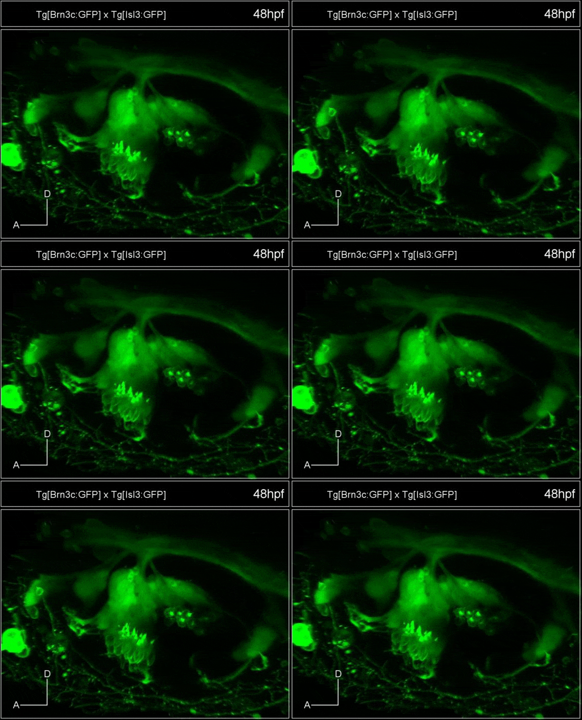
Figure 1: The sensory organ of the inner ear. Inner ear of a zebrafish embryo in which ciliated cells (hair-like appearance), organized in two sensory patches, and neurons of the sensory ganglion in contact with the hair cells on one side and on the other sending projections with information to the brain.
Figure 2: Following the progenitor cells in real time. Left: History of the coloured progenitor cells (see legend) considering the moment at which they delaminate from the otic vesicle. Early (white) delamination cells are separated from those that have arrived late (red) once in the neural ganglion. Right: Coloured progenitors attending to their position in the otic vesicle at the time of delamination (blue front and red back). The cells maintain their relative position in the ganglion (blue and red do not mix).
