Image gallery
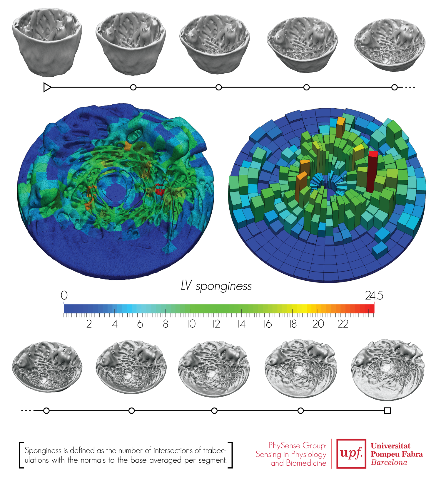
Figure 1: Subject independent reference frame for the left ventricular detailed cardiac anatomy and the evaluation of left ventricular sponginess.
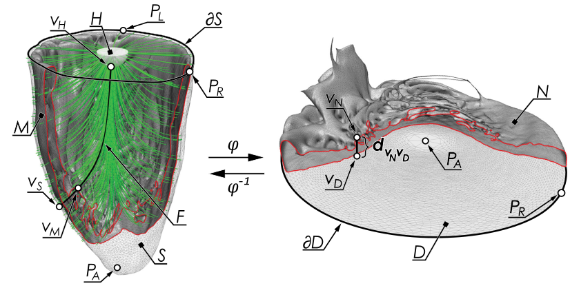
Figure 2: Illustration of a mapping procedure of a detailed anatomical LV mesh to the patient independent reference frame.
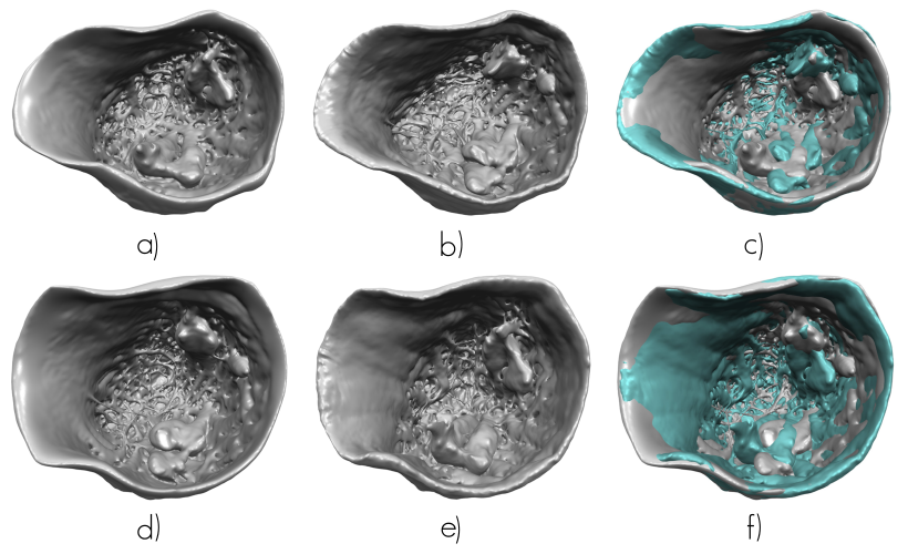
Figure 3: Illustration of LV of two datasets including inflow and outflow tracts, and them mapped onto each other’s bounding surface. a) Original HH 84 LV mesh. b) Mesh HH 88 LV mapped onto HH 84 LV bounding surface. c) Overlap of a) (gray color) and b) (turquoise color). d) Original HH 88 LV mesh. e) Mesh HH 84 LV mapped onto HH 88 LV bounding surface. f) Overlay of of d) (gray color) and e) (turquoise color).
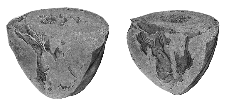
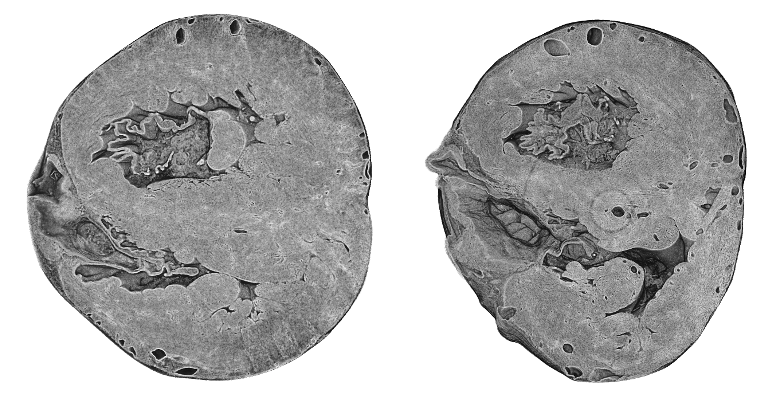
Figure 4: Visualization of normal and intrauterine growth restricted rabbit hearts acquired by X-ray phase-contrast synchrotron radiation-based micro-CT.
Figure 7: Ultrasound imaging – three methods to extract motion information from 4 chamber view acquisitions. 1) Fixed sample: reads motion information from myocardial velocity imaging. A user-defined region of interest (ROI) is kept static throughout the heart cycle to maintain the analysis and the interaction as simple and reproducible as possible. 2) Manual tracking: reads motion information from myocardial velocity imaging. It requires the user to define a ROI on the myocardium at end-systole and end-diastole. This ROI is interpolated in space to mimic the myocardial movement. 3) Speckle tracking: uses block matching techniques to follow speckles from B-mode images and estimate the myocardial motion.