
Cochlear implants
High resolution statistical shape models of the cochlea
In order to better understand the shape and function of the inner ear, and aiming at the creation of computational models for surgical planning and simulation, we have worked on the analysis of cadaveric high resolution microCT datasets. These data protray minute details that are not visible in routine clinical CT or MRI scans performed for diagnosis and surgical planning.
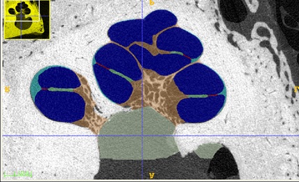
Segmentation of high-resolution microCT images of the inner ear based on Random Walks (Ruiz et al., International Journal of Computer Assisted Radiology and Surgery 2016; Ruiz et al., Image and Vision Computing 2016)
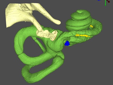
Applying these segmentation methods to multiple datasets, it is possible to build a Statistical Shape Model (SSM) representing the average shape and the principal modes of variation, i.e. in which predominant ways the shape varies across the population. Recent work has focused on the construction of these statistical models, notably on the algorithms for correspondence establishment, needed to align all datasets prior to the computation of the SSM. Furthermore, the SSM has been incorporated into a segmentation algorithm, effectively leading to the estimation of patient-specific high-detail cochlear shape from low resolution clinical data.
SSM model construction, including Laplacian-based correspondence establishment (Kjer et al., Pattern Recognition Letters 2016)
Surgical planning for cochlear implantation
Cochlear implantation involves the insertion of a thin electrode array inside the cochlea. This is a very delicate procedure, and its success is key to the intervention. Furthermore, there is growing evidence stating that the insertion depth of the electrode array has a strong influence on the quality of perceived sound after implantation. We have developed a system to perform patient-specific simulations of electrode array insertion, based on the detailed anatomy of the patient (estimated thanks to the SSM) and a library of implants available in the market. A planning software has been developed to assist the surgeon in planning the intervention and exploring different choices of implant and surgical parameters.
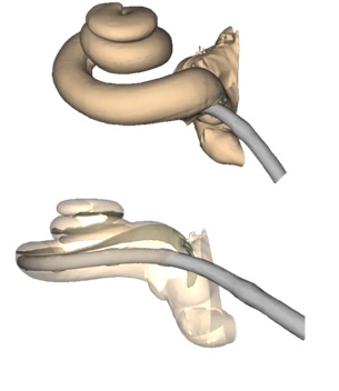
Virtual electrode array insertion, for the automatic construction of patient-specific finite element models for simulation (Mangado et al., Annals of Biomedical Engineering 2016)
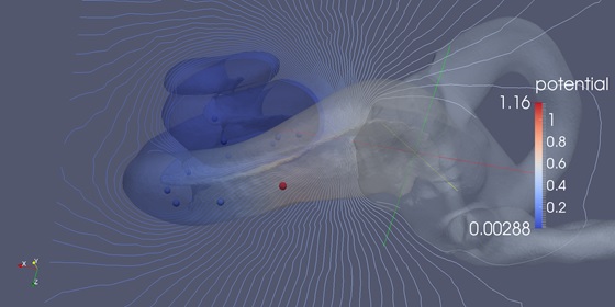
Computational simulation of cochlear implants
Our automatic framework is able to generate patient-specific models for subsequent finite element simulations. In particular, we can simulate the activation of the cochlear implant in different configurations and using specific activation protocols. The electrical distribution around the cochlea is computed, and this is coupled with a model of the nerve fibres around the cochlea. The action potentials activating these nerves are evaluated, effectively assessing neural function and therefore sound perception. Work in collaboration with NASA Ames Research Center has focused on the simulation of the effect of nerve degeneration on their electrical activation, in the particular context of cochlear implants.
Simulation of electrical stimulation protocols for cochlear implants (Ceresa et al., Molecular Neurobiology 2015)