
CRG/UPF Advanced Light Microscopy Unit (ALMU)
Nadia Halidi (Head of the Advanced Light Microscopy Unit)
Website
Research Outline
Our unit serves as a core facility for high-end light microscopy for PRBB researchers. A range of instruments with unique capabilities fully covers the spectrum of advanced imaging applications. We provide advice in the initial experiment planning, training of the researchers on the instruments and assistance with the subsequent data analysis. In 2018, the total booked microscope usage time of the unit reached more than 19000 hours in more than 6300 separate bookings. During the year, a total of 216 researchers from PRBB and one company used the unit, as well as projects from external visitors. On average, 94 investigators use the unit every month. During the first months of 2018 the unit finished moving into its new location in the basement of the PRBB building. The instrumentation of the unit was kept up to date by targeted upgrades of the existing equipment.
Current Projects
The unit is currently developing transient darkstate imaging protocols for the fast quantitative assessment of fluorophore blinking and the optimization buffers for super-resolution imaging (see figure).
Timo Zimmermann is the Spanish representative for Biological Imaging in the Interim Board of the ESFRI project Euro-BioImaging. The microscopy unit participates in two highly evaluated Spanish node proposals for Euro-BioImaging, for super-resolution microscopy (with the Photonic Sciences Institute ICFO) and for in-vivo and intravital imaging (with the University of Barcelona and IRB Barcelona) which are participating in the interim operation phase of Euro-BioImaging which has started in May 2016.
The unit participated in the execution of BIST Winter School on Microscopy and Imaging Sciences 2018 which took place from January to February for the BIST Master Course of Multidisciplinary Research in Experimental Sciences.
Team during 2017-18
- Technicians:Xavier Sanjuan (UPF), Raquel García (CRG), Arrate Mallabiabarrena (CRG), Raúl Gómez (CRG).
-
Research visits: Cecícia Rotondaro, Personal de Apoyo Científico, IIBBA, Fundación Instituto Leloir, Argentina. Dates of visit: September-November 2016.
Selected publications 2017-18
- Bosch-Presegué L, Raurell-Vila H, Thackray JK, González J, Casal C, Kane-Goldsmith N, Vizoso M, Brown JP, Gómez A, Ausió J, Zimmermann T, Esteller M, Schotta G, Singh PB, Serrano L, Vaquero A (2017) Cell Rep. 21(8):2048-2057 [doi: 10.1016/j.celrep.2017.10.092].
- Merino D, Mallabiabarrena A, Andilla J, Artigas D, Zimmermann T, Loza-Álvarez P (2017) STED Imaging Performance Estimation By Means Of Fourier Transform Analysis Biomed. Opt. Express 8 2472-2482.. F.I.:3.337 [doi:10.1364/BOE.8.002472].
- Raote I, Ortega Bellido M, Pirozzi M, Zhang C, Melville D, Parashuraman S, Ximmermann T, Malhotra V (2017) TANGO1 Assembles Into Rings Around COP II Coats At ER Exit Sites J. Cell Biol. 216 901-909.. F.I.:7.955 [doi:10.1083/jcb.201608080].
Other relevant information 2017-18
Timo Zimmermann, Invited Speaker in the Spanish Portuguese Meeting for Advanced Optical Microscopy (SPAOM), Granada, 24-26th October 2018. Title: “How to get to know your fluorophore in less than a millisecond”.
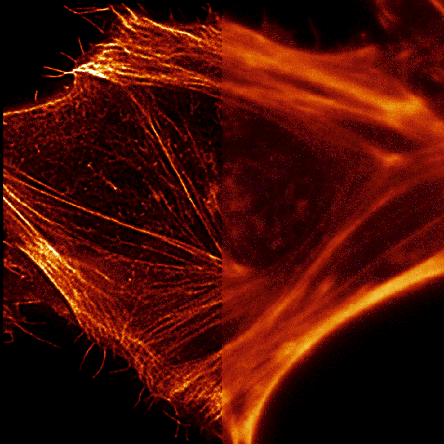
Total Internal Reflection Fluorescence (TIRF) image of a fixed Hela cell stained with Silicon-Rhodamin (SiR) Actin to visualize the actin cytoskeleton. The resolution of the image based on the average collected fluorescence of the image series (right) can be significantly improved by analysing local fluorescence fluctuations between images using the Super-Resolution by Radial Fluctuation (SRRF) algorithm (left). Strong transient darkstates were induced in the SiR Actin label by imaging the sample in a buffer in which oxygen was removed by glucose oxidase and 1 mM ascorbic acid was added.