QCM, an advanced technique to process cardiac imaging data
QCM, an advanced technique to process cardiac imaging data
A new method described in a study by the PhySense research group in conjunction with the Arrhythmia Section of the Cardiology Service at Hospital Clínic, Barcelona, published in Medical Image Analysis.
A study published on 25 March in the advanced edition of the journal Medical Image Analysis by researchers of the PhySense research group of the Department of Information and Communication Technologies (DTIC) (D. Soto, C. Butakoff and O. Cámara ), in conjunction with the Arrhythmia Section of the Cardiology Service of Hospital Clínic, Barcelona, presents a new, more accurate technique to integrate complementary information about the heart, to guide interventions required by ventricular tachycardia patients who have suffered a heart attack.

In a heart attack, the lack of blood supply to heart tissue causes the affected area to cease to function and progressively form a scar. Although magnetic resonance imaging (MRI) provides information on those areas of the heart in which muscle tissue is not functional, the necrotic tissue of the scar is not entirely homogeneous, that is, it presents intermediate areas that are still functional that are the ones that give rise to many of the ventricular tachycardia observed, given that they generate abnormal electrical circuits (reentries).
In these cases some of the most advanced medical intervention is radiofrequency ablation of intermediate areas of the scar in order to generate short circuits and stop ventricular tachycardia. In the course of heart scar tissue ablation an electroanatomical system is used that provides additional information about the heart to discern between healthy tissue, which conducts electrical propagation normally, from sick tissue, with slower or non-existent conduction.
Integrating the information is crucial to guide the intervention
In order to guide radio frequency ablation, the doctor must be able to combine the information available both from MRI images and from the electroanatomical system. This integration can also be done semi-automatically using advanced computational image processing methods.
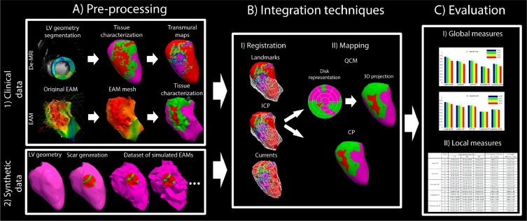
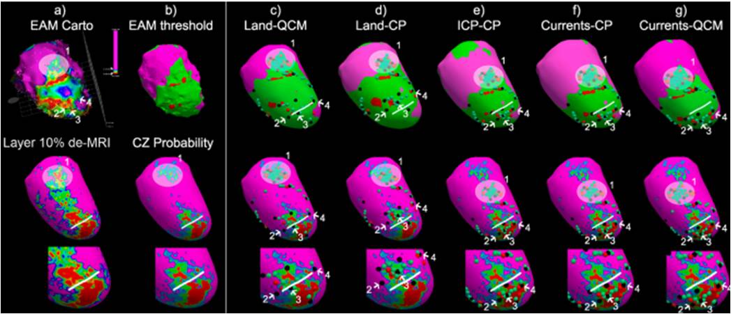
The study published in Medical Image Analysis presents the Quasi-Conformal Mapping (QCM) method to perform the integration of information about the scar from the MR images and electroanatomical systems, and it is compared with different state-of-the-art strategies by means of controlled synthetic data and a complete set of clinical data.
The QCM method is more accurate in integrating data
QCM technology facilitates data integration via the homeomorphic projection of the three-dimensional geometry of the heart ventricles to a two-dimensional space. The authors of the study have confirmed the validity of this method in comparison with other existing methods, and the result has been that QCM has proven to be more accurate in integrating multimodal data than other available methods, both with synthetic and clinical data.

Close technological, clinical and entrepreneurial collaboration
It is important to mention that in order to ensure that the computational tools developed by the PhySense research group are appropriate for clinical environments, the researchers have worked very closely with Dr. Antonio Berruezo Sánchez, a member of the Arrhythmias Section of Hospital Clínic, Barcelona.
As a result of this relationship, David Soto, a researcher of the PhySense research group and first author of the study, was recently hired as a research engineer at the Arrhythmias Section of Hospital Clínic, Barcelona, partially to work in the clinical evaluation of the methodology described in this study, which is the subject of the doctoral thesis he plans to read at the Department of Information and Communication Technologies (DTIC) at UPF at the end of 2016.
Óscar Cámara, principal investigator of the study noted that “for the future, in the event of having a positive clinical evaluation in a sufficient number of cases, QCM could become part of the software for viewing and processing medical data for arrhythmias”. Such software is currently being developed jointly by Hospital Clínic de Barcelona with the company Galgo Medical, with the collaboration of Pompeu Fabra University.
Reference work:
David-Soto Iglesias, Constantine Butakoff, David Andreu, Juan Fernández Armenta, Antonio Berruezo, Óscar Cámara (2016), “Integration of electro-anatomical and imaging data of the left ventricle: An evaluation framework”, Medical Image Analysis, vol. 32, pp. 131-144.
