A study by UPF and ULiège reveals new insights into consciousness, which establish the basis for treating disorders such as vegetative state in the future
A study by UPF and ULiège reveals new insights into consciousness, which establish the basis for treating disorders such as vegetative state in the future
Currently, the diagnosis of people with consciousness disorders is mainly based on their behavioural response to external stimuli. However, this study examines the underlying brain activity of people with these disorders, laying the foundations for improving their diagnosis and treatment in the future. The research used simulations of brain functioning with a computer model.
A neuroscientific study has revealed new insights into consciousness mechanisms, which establish the basis for treating disorders such as so-called vegetative state in the future. The study is part of the Human Brain Project and has been jointly conducted by researchers from UPF and the University of Liège (ULiège) in Belgium. The results have recently been published in the journal Human Brain Mapping, in the article “Whole-brain analyses indicate the impairment of posterior integration and thalamo-frontotemporal broadcasting in disorders of consciousness”.
The results of the study could change and improve the way in which disorders of consciousness are diagnosed today. Currently, patient diagnosis is mainly based on their behavioural response to external stimuli provided by medical professionals, but this study focuses on examining the underlying brain activity of people with these disorders (not- or not necessarily - linked to environmental stimuli).
Jitka Annen, principal investigator (ULiège): “the physician sits down with the patient and assesses their response to stimuli. However, this may not correspond to their underlying brain activity: some patients with high underlying activity may still be unable to react [...] We wanted to go one step beyond”
Jitka Annen, principal investigator of the study (ULiège) explains: “The physician sits down with the patient and assesses their response to stimuli. However, this may not correspond to their underlying brain activity: some patients with high underlying activity may still be unable to react. It is a heterogeneous group. We wanted to go one step beyond neuroimaging-based assessment and classification and look at the information flow in their brains, to find common patterns associated with consciousness”.
Gorka Zamora-López, also principal investigator of the study from the UPF Center for Brain and Cognition (CBC), clarifies that the results of this study could lead to more accurate diagnoses than those currently obtained from medical tests based on external stimuli. It could be the case that a patient does not respond to external stimuli due to factors other than vegetative state (for example, because of mobility problems, hearing deficit…). If the diagnosis is not made on the basis of external stimuli, but on the basis of the underlying brain activity, it will be more accurate in avoiding situations that may give rise to error.
The brain activity of patients with different disorders of consciousness is studied: vegetative state and minimally conscious state
Currently, patients with DoC (Disorders of Consciousness) are classified into several categories: coma, unresponsive wakefulness syndrome (formerly known as vegetative state), and minimally conscious state (which has traditionally been defined as the state in which a person begins to partially respond to external or environmental stimuli).
The researchers analysed two groups of DoC: unresponsive wakefulness syndrome and minimally conscious state, along with a control group of healthy people. These analyses were carried out both with real people and with simulations of brain functioning using a computer model.
Testing brain dynamics with real patients
In the case of tests with real people (healthy, in vegetative state, and in minimally conscious state), data were collected on their brain dynamics in resting state (awake, but without any assigned task) by means of fMRI (functional magnetic resonance imaging). In these analyses, spontaneous disturbances in brain activity were observed and the likelihood of them moving from one part of the brain to another.
“Based on spontaneous spikes in activity, we evaluate the personalized connectivity of each patient’s brain, which can tell us how likely it is that a signal will travel from one point to another”, explains CBC-UPF’s Gorka Zamora-López, principal investigator of the study.
Simulations of brain functioning with a computer model
The researchers of the Human Brain Project have also used an approach based on simulations of brain functioning using a computer model to work to identify the brain circuits involved in consciousness. They studied the broadcasting of signals in models of the brain of patients with disorders of consciousness (DoC) and identified two relevant circuits in the posterior cortical region and the thalamus and frontotemporal region.
These simulations have served to analyse which regions of the brain are more likely to broadcast signals and which regions to receive them in different types of people (healthy, in vegetative state and in minimally conscious state).
“After building an individualized computational model of the patient, we activated a signal in the model and observed how the brain reacted. In particular, we looked for which areas were most likely to respond to a signal; which areas are most likely to broadcast it. Basically, we look at whether an area acts as influencing or as influenced” – Gorka Zamora-López (CBC-UPF) clarifies.
The results of the simulation of brain functioning
The following image shows the results of this simulation. In the first column, the results are shown for a healthy person; in the second, for a person with unresponsive wakefulness syndrome (or vegetative state); and in the third, for a person in minimally conscious state. The part above the dashed line shows the regions of the brain most likely to broadcast, and the lower part, the most likely to receive signals.
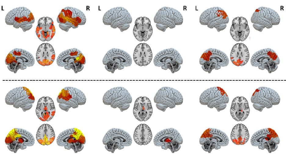
These results show a marked distinction between the unresponsive wakefulness syndrome group and the minimally conscious state group, since the former shows no activity in the identified brain circuits. “The key difference is that, in patients with unresponsive wakefulness syndrome, no region of the brain appears embedded in a functional network, all show equally low activation. In contrast, different regions and circuits appear in the brain models of people in minimally conscious state: the thalamo-frontotemporal region when it broadcasts signals, and the posterior cortical region when it receives them”, adds Rajanikant Panda of the University of Liège.
Gorka Zamora-López (UPF-CBC): “I think these results could guide physicians to better understand what is failing in information exchange and thus look for ways to reactivate these circuits”
These findings provide new insights into disorders of consciousness. They could lead to a more accurate understanding of the mechanisms of consciousness, based on brain activity and not on behavioural responses. “I think these results could guide physicians to better understand what is failing in information exchange and thus look for ways to reactivate these circuits”, Zamora-López (UPF) concludes.
Based on the text by Roberto Inchingolo
Reference article:
Rajanikant Panda, Ane López-González, Matthieu Gilson, Olivia Gosseries, Aurore Thibaut, Gianluca Frasso, Benedetta Cecconi, Anira Escrichs, Coma Science Group Collaborators, Gustavo Deco, Steven Laureys, Gorka Zamora-López, Jitka Annen. Whole-brain analyses indicate the impairment of posterior integration and thalamo-frontotemporal broadcasting in disorders of consciousness. Human Brain Mapping. https://doi.org/10.1002/hbm.26386