Towards a better understanding of the cellular architecture of the human brain
Towards a better understanding of the cellular architecture of the human brain
According to a study published on January 9 in the journal Science Advances, led by the National University of Singapore, co-authored by Gustavo Deco, ICREA research professor and director of the Center for Cognition and Brain.

An inter-disciplinary research team led by scientists from the National University of Singapore (NUS) has successfully employed machine learning to uncover new insights into the cellular architecture of the human brain.
The team demonstrated an approach that automatically estimates parameters of the brain using data collected from functional magnetic resonance imaging (fMRI), enabling neuroscientists to infer the cellular properties of different brain regions without probing the brain using surgical means. This approach could potentially be used to assess treatment of neurological disorders, and to develop new therapies.
The work was published on January 9 in the journal Science Advances and has had the collaboration of Gustavo Deco, research professor ICREA and director of the Center of Cognition and Brain CBC of the Department of Information Technologies and Communications DTIC of the Pompeu Fabra University.
"This work means a quantum leap in that we also incorporate heterogeneity in the local dynamics of each node, that is, we optimize them in order to obtain a much more reliable and realistic biophysical and physiological model"
As Professor Gustavo Deco has explained: "Until now, in general, the models of the whole brain were limited by the fact that the local dynamics of each node or brain region was identical, that is, homogeneous. In this way, through the heterogeneity of the anatomy extracted through techniques of Diffusion Tensor Imaging (DTI) and tractography, many interesting aspects of the functional dynamics of the brain were explained. " "However, in this new work we make a quantum leap in that we also incorporate heterogeneity in the local dynamics of each node, that is, we optimize them in order to obtain a much more reliable and realistic biophysical and physiological model", adds Deco, co-author of the study.
The underlying pathways of many diseases occur at the cellular level, and many pharmaceuticals operate at the microscale level. To know what really happens at the innermost levels of the human brain, it is crucial for us to develop methods that can delve into the depths of the brain non-invasively.
The new study, conducted in collaboration with researchers from the Netherlands and Spain was first reported online in scientific journal Science Advances on 9 January 2019.
Unravelling the complexity of the human brain
The brain is the most intricate organ of the human body, and it is made up of 100 billion nerve cells that are in turn connected to around 1,000 others. Any damage or disease affecting even the smallest part of the brain could lead to severe impairment.
Currently, most human brain studies are limited to non-invasive approaches, such as magnetic resonance imaging (MRI). This limits the examination of the human brain at the cellular level, which may offer novel insights into the development, and potential treatment, of various neurological diseases.
Different research teams around the world have harnessed biophysical modelling to bridge this gap between non-invasive imaging and cellular understanding of the human brain. The biophysical brain models could be used to simulate brain activity, enabling neuroscientists to gain insights into the brain. However, many of these models rely on overly simplistic assumptions, such as, all brain regions have the same cellular properties, which scientists have known to be false for more than 100 years.
Constructing virtual brain models
Asst Prof Yeo and his team worked with researchers from Universitat Pompeu Fabra, Universitat Barcelona and University Medical Center Utrecht to analyse imaging data from 452 participants of the Human Connectome Project. Departing from previous modelling work, they allowed each brain region to have distinct cellular properties and exploited machine learning algorithms to automatically estimate the model parameters.
This form of hierarchical processing is a key feature of both the human brain and recent advances in artificial intelligence.
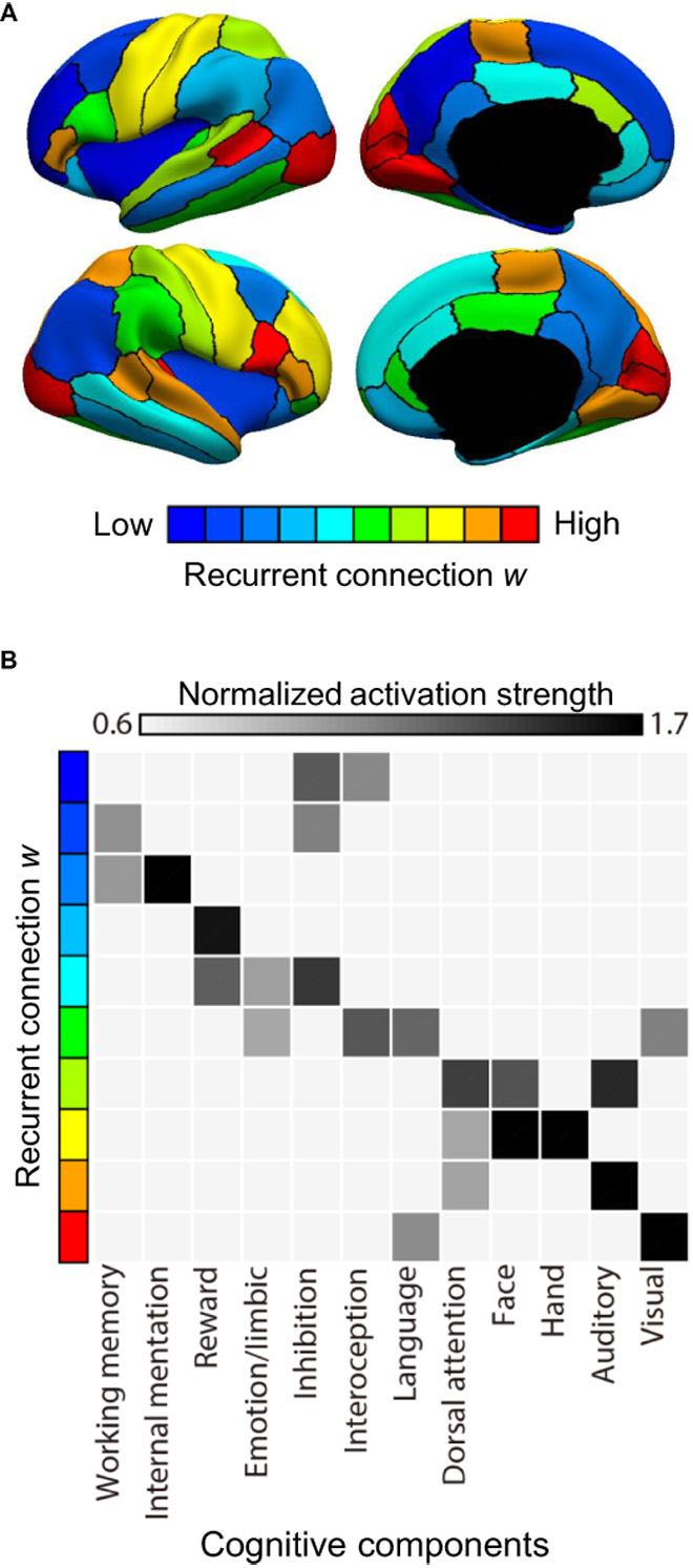
The research team found that brain regions involved in sensory perception, such as vision, hearing and touch, exhibit cellular properties opposite from brain regions involved in internal thought and memories. The spatial pattern of the human brain's cellular architecture closely reflects how the brain hierarchically processes information from the surroundings. This form of hierarchical processing is a key feature of both the human brain and recent advances in artificial intelligence.
This study suggests that the processing hierarchy of the brain is supported by micro-scale differentiation among its regions, which may provide further clues for breakthroughs in artificial intelligence.
Moving forward, the NUS team plans to apply their approach to examine the brain data of individual participants, to better understand how individual variation in the brain's cellular architecture may relate to differences in cognitive abilities. The team hopes that these latest results can be a step towards the development of individualised treatment plans with specific drugs or brain stimulation strategies.
Related work:
Peng Wang, Ru Kong, Xiaolu Kong, Raphaël Liégeois, Csaba Orban, Gustavo Deco, Martijn P. van den Heuvel, BT Thomas Yeo (2019),"Inversióof a large-scale circuit model reveals a cortical hierarchy in the dynamic resting human brain", 9 January, vol. 5, number 1, eaat7854, DOI: 10.1126 / sciadv.aat7854.