Researchers identify a fetal biomarker for the prognosis of brain abnormalities
Researchers identify a fetal biomarker for the prognosis of brain abnormalities
They are studying ventriculomegaly, a condition associated with the appearance of brain abnormalities in the long term. The results of the study have been published in the journal NeuroImage: Clinical and its authors are members of the SIMBIOsys research group, together with researchers of Hospital Clínic and Hospital Sant Joan de Déu in Barcelona.
Ventriculomegaly is a condition that occurs 0.3-1.5 times in every 1,000 new-borns and involves an increase in the size of one of the two lateral ventricles, cavities located inside each of the cerebral hemispheres, where the cerebrospinal fluid is produced that protects the central nervous system: the brain and the spinal cord.
There is some relationship between the degree of dilation of the lateral ventricles with the development of the central nervous system and the onset of brain abnormalities in the long term. The main aim of the research is the study of isolated non-severe ventriculomegaly and its relationship with cortical folding and the results were published in open access on 28 January in the online edition of the journal NeuroImage: Clinical.
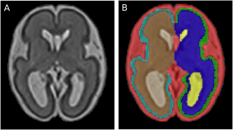
The study of the brain through imaging techniques plays a crucial role in understanding brain abnormalities and early diagnosis. It is essential to study brain abnormalities in utero and the evaluation of deviations in the event of maldevelopment. In this work, brain magnetic resonance images were used from 23 human fetuses with a diagnosis of isolated non-severe ventriculomegaly and 25 healthy controls, between 26 and 29 weeks of gestation to identify deviations of the folding of the cerebral cortex related to ventriculomegaly and neurological development.
The results of the study indicate that there is a decreased cortical folding in fetuses with ventriculomegaly. More specifically, a significant cortical folding decrease in the insula, the posterior part of the temporal lobe and occipital lobe. As it is known that there is a high risk of neurological damage to fetuses presenting ventriculomegaly, a condition that may be related to other neurodevelopmental processes, this study shows that cortical regions with altered folding could constitute good potential biomarkers for the prognosis of brain abnormalities and ventriculomegaly is an indicator of altered cortical development.
The authors of this study are members of Pompeu Fabra University linked to the SIMBIOsys (Simulation, Imaging and Modelling for Biomedical Systems) research group of BCN MedTech, including Miguel Ángel González Ballester, ICREA research professor with the Department of Information and Communication Technologies (DTIC) and group director, together with Gemma Piella, Oualid M. Benkarim and Gerard Sanroma, who have carried out the research with Eduard Gratacós, Elisenda Eixarch and Nadine Hahner, researchers of Hospital Clínic and Hospital Sant Joan de Déu in Barcelona and members of the network on rare diseases CIBER-ER.
Reference work:
Oualid M. Benkarim, Nadine Hahner, Gemma Piella, Eduard Gratacós, Miguel Ángel González Ballester, Elisenda Eixarch, Gerard Sanroma (2018), “Cortical folding alterations in fetuses with isolated non-severe ventriculomegaly”, Neuroimage:Clinical, Volume 18, pp. 103–114. https://doi.org/10.1016/j.nicl.2018.01.006. Open access.
Image, Figure 2 of the article: Fetal brain MRI segmentation: 26.4 GW-old fetus with right INSVM (A) and corresponding segmentation (B), with different labels for left and right WM, cortex and lateral ventricles