The results of the European project HEAR-EU are published in open access in the journal Nature Scientific Data
The results of the European project HEAR-EU are published in open access in the journal Nature Scientific Data
The results of the European project HEAR-EU are published in open access in the journal Nature Scientific Data
Thus the main objective of the project, to develop computer systems from high resolution computer assisted microtomography images of the human inner ear for the design of cochlear implants and support for surgery has been achieved. It was coordinated by Miguel A. González Ballester, ICREA research professor and director of the SiMBioSys Research Group, BCN Medtech Research Unit.
The results of the European project of the EU’s 7th Framework Programme, HEAR-EU, are presented in the high impact factor journal Nature Scientific Data and are available to professionals in open access. The project has received funding of 4.7 million euros and was carried out from 2012 to 2015.
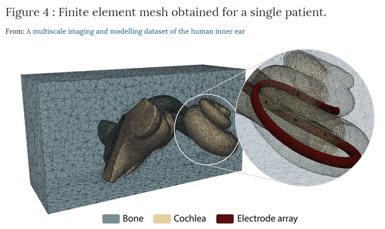
The main goal of HEAR-EU was to develop computer systems based on computer-assisted microtomography, that is to say, high-resolution 3D X-ray imaging (microCT), and predictive mathematical models for the design of cochlear implants and to support surgery of the inner ear. The images and the information obtained will enable better planning of surgical interventions in a personalized way, making them less invasive and improving the design of implants.
So, a most important resource for both biomedical and computer engineers working on the design of electrodes and auditory implants, such as surgeons and specialist physicians who need to have precise anatomical knowledge of the internal structures of the body, is being made available in open access.
Miguel A. González Ballester, ICREA research professor and coordinator of the SiMBioSys (Simulation, Imaging and Modelling for Biomedical Systems) Research Group, part of the BCN Medtech research unit at the Department of Information and Communication Technologies (DTIC) at UPF, coordinated this project. The consortium was composed of three academic partners: Pompeu Fabra University, the University of Bern (Switzerland), and the Technical University of Denmark; together with two SMEs: Alma IT Systems and SCANCO Medical, and one multinational company, MED-EL GmbH.
This collection of high-resolution data of the human inner ear was carried out by acquiring cone beam computer tomography (CBCT) and X-ray computer tomography to achieve cross sections of physical objects that can be used to recreate a virtual model (3D model) without destroying the original object, which together with manual delineations of the human cochlea, has made it possible to obtain a statistical model that codes the human anatomical variability of this organ and provides invaluable information for the insertion of electrodes and electrical and functional simulations to predict the recovery of hearing as a result of implantation.
This new resource is a valuable methodological tool for research and for all those professionals, computer scientists, engineers, and clinicians working in the different fields of image analysis, computer simulations, medical imaging and biomedical engineers designing new strategies to obtain cochlear implants and other medical devices.
Reference work:
Nicolas Gerber, Mauricio Reyes, Livia Barazzetti, Hans Martin Kjer, Sergio Vera, Martin Stauber, Pavel Mistrik, Mario Ceresa, Nerea Mangado, Wilhelm Wimmer, Thomas Stark, Rasmus R. Paulsen, Stefan Weber, Marco Caversaccio & Miguel A. González Ballester, (2017), “A multiscale imaging and modelling dataset of the human inner ear”, Nature Scientific Data, published online, 19 September.